1. Budova and functions nervous system... Glia.
2. Reflex. Reflex arc. Classification of reflexes.
3. Vіkovі features of the brain and spinal cord.
1. Budova and functions of the nervous system. glia
The nervous system regulates and coordinates the activity of the organisms and systems, which enhances the function of the organisms. The bosses of the їy zdіysnyuє link to the body with a new middle-class and th-th adaptation to the progressively young minds. The nervous system is the material basis of the intelligence of people, their misery, behavior, mov.
The brain and spinal cord are brought to the central nervous system. The offense of stench is evolutive, morphological and functionally tied between oneself and without a sharp separation to go one into one.
Function of the nervous system
1. Zabezpu links to the body with a zonishnim middle.
2. I will secure the interconnection of all parts of the body between myself.
3. For the regulation of trophic functions, to regulate the exchange of words.
4. Nervous system, brain sprout, є a substrate of mental activity.
Functionally, the nervous system is susceptible to somatic and autonomous (autonomic), anatomically - to the central nervous system and peripheral nervous system
The somatic nervous system regulates the skeletal muscles and ensures the sensitivity of the human body. An autonomous (vegetative) nervous system regulates the exchange of words, the robot of internal organs and smooth cells.
The vegetative nervous system is the nerve system for all internal organisms. Vona will also preserve the trophic innervations of skeletal tissues, organs and tissues, and the nervous system itself.
The peripheral nervous system is established by the numerical male nerves, nerve gossips and nodes. Nerves deliver impulses from the central nervous system directly to the working organ - the muscles - information from the periphery in the central nervous system.
The main elements of the nervous system are cells of the nerves (neurons). The confirmation of the clerical theory of the budov and the nervous system was taken into account behind the supplementary electronic microscopy, which showed that the membrane of the nerve cells is not the main membrane of the young cells. It seems to be susceptible to the entire surface of the nerve cells and the emerging cells of the cells. Skin nerve cells are anatomically, genetically and metabolic units, as well as cells and tissues of the body. People's nervous systems have close to 100 billion nerve cells... Fragments of the dermal nerve cord is functionally tied to thousands of neurons, a number of many options such sounds are very close to ness. Nervous cells should be able to see as one of the analogs of the organisation of the nervous system, of the full molecular, synaptic, subclinics of the supracellular lines of the canal neural lines, of the nervous centers and of the behavioral systems.
Budova neuron. The thickness of the neuron, which is tied with sprouts, є the central part of the neuron, and will prevent the harvesting of the central part of the cell. Tilo in with a sharuvate membrane, which are two balls of lipids with a protolezhny arrangement, to set the matrix, in which the bricks are inserted. The thickness of the neuron is the nucleus or the nucleus, which can take revenge on the genetic material.
The nucleus regulates the synthesis of proteins in all cells and controls the differentiation of young nerve cells. The cytoplasm of the neuron has a large number of ribosomes. Some ribosomes grow vividly in the cytoplasm by one or the purchase. The Іnshi ribosomes are attached to the endoplasmic reticulum, which is the internal system of membranes, tubules, bulbs. Attached to the ribosome membranes and synthesized beads, which can then be transported from the cells. The purchase of the endoplasmic reticulum from the ribosomes embedded in it becomes characteristic of those neurons, the substance of Nisl. Purchase of smooth endoplasmic reticulum, in which ribosomes are not inserted, store the Golgi apparatus; to transfer, which is important for the secretion of neurotransmitters and neuromodulators. Lyzosomes are a storage in the membrane of the purchased hydrolytic enzymes. Important organelles of nerve cells - mitochondria - are the main structures of energy supply. On the inner membrane of the mitochondria, there is all the enzyme in the citric acid cycle - the most effective way to break down glucose, which is dozens of times more effective than anaerobic way. In nerve cells, there are also microtubules, neurofeatures and microfilmings, which grow in diameter. Microtubules (diameter 300 nm) pass through the nerve cell into the axon and dendrites, and are an internal transport system. Neurofilment (diameter 100 nm) is seen only in the nerve cells, especially in the great axons, and also store a part її transport systems... Mikrofilamenti (diameter 50 nm) bends well in the growths of nerve cells, the stench takes a part in some types of interneuronal diseases.
Dendrites are tree-like outgrowths of a neuron, its head is not receptive field, so that information is not provided, as it comes through synapses from all neurons or directly from the middle. When the dendrites are visible, the dendrites are reddened: the number of dendritic gaps grows, and the diameter of the dendrites sounds. On the surface of the dendrites of the bagatoch neurons (pyramid neurons of measles, cells of the Purkin's moss and іn.) Є spines. The spine apparatus is a storage part of the dendrite canaliculi system: microtubules, neurofilments, Golgi apparatus and ribosomes are located near the dendrites. Functional maturation and ear of active activity of nerve cells to grow with the appearance of spines; Trivial to pinpoint the appropriate information to the neuron until the spines are removed. The appearance of thorns is larger than the surface of the dendrites.
Axon є for a single person, call up the spine of a neuron, a service for quick implementation of the neuron. In the beginning, you can go on a bike (up to 1000), a number of grips.
The nerves of the clitini display a number of out-of-the-box functions, which are directed to the control of the power processes of the organization. Tse of exchange of words with navkolishnіm middle, Osvita and vitrachnya energy, synthesis of bilky and in. In addition, the nerves of the clientele are aware of the authorities only and specific functions for the reception, reprocessing and collection of information. Neurons create information, replay (code) information, quickly transmit information on specific routes, organize communication with other nervous cells, secure information and generate information. For the identification of these functions, neurons may polarize organization from the bottom of the inputs and outputs and to replace a number of structural and functional parts.
Classification of neurons. Neurons move on the onset of groups: according to the mediator, they can be seen in the end of axons, develop adrenergic, cholinergic, serotonergic neurons, etc.
Neurons of the somatic and autonomic nervous systems are clearly seen from the central nervous system.
Directly informatization of the onset of neurons:
Affection, how to use additional receptors for information about the development and the internal medium of the body and transmit it to the central nervous system;
Effective, how to transmit information to the working organs - effectors (nerves of the cells, who are responsible for the performance, sometimes they are called effectors);
Inserts (interneurons), which provide for the interaction between the neurons of the central nervous system.
Behind the inflow, they see the zbudzhuyut and galvanize neurons. For the activity of developing background activity and "moving" neurons, so that they can only be generated in response to development. The background activity of the neurons is perceived as a distant small of the generation of impulses, so as one neuron is discharged without interruption (rhythmically or arrhythmically), and in bursts of impulses. The interval between pulses in pachts becomes milliseconds, between batches - seconds. Background neurons play an important role in improving the tone of the central nervous system, especially measles in the brain.
To use the sensory information of the neuron to be mono- and bipolysensory. Monosensory neurons to the center of hearing in the cortex of the great brain. Bisensory neurons are detected in the secondary zones of the analyzer in the cortex (the neurons of the secondary zone of the healthy analyzer in the cortex of the great brain react to light and sound stimuli). Polysensory neuron - tse neurons of the associative zones of the brain, motor measles; the stench reacts to the development of receptors in school, healthy, auditory and other analysators.
The nerves of the cells are tied together by numerical links: the nerves of the axon of one neuron stick to the dendrites of the other neuron, or the distribution of the axon surrounds all the small of the neuron. The point of contact of neurons is called synapses.
Synapse is a structural statement, which prevents the transfer of energy from the nerve cell to the nerve cell, or from the nerve cell to the cell of the robotic organ. The term "synapse" was pro-proponated by the English physiologist Ch. Sherrington.
Whether the synapse is composed of 3 parts - the presynaptic widdil, the synaptic weddil and the postsynaptic widdil.
The presynaptic part is folded from the end of the axon, covered by the presynaptic membrane. In the middle there are bulbs - vesicles cheery speech- mediator.
The synaptic gap is stored with a line, close behind the warehouse to the blood plasma.
The postsynaptic form of representations by the postsynaptic membrane, in which there are chemoreceptors, are sensitive to the singing mediators.
The synapse has a great number of mitochondria.
The electrical impulse is energized, moving along the axon, reaching the synaptic chicks, as a result of which the formation and development will take place. Acetylcholine is introduced into the bulb, which, through the pores of the presynaptic membrane, enters the synaptic cleft and enters into the chemical interaction with the receptors of the postsynaptic membrane. As a result, the collapse of the cations in calories and the collapse of the cations in sodium significantly increase, the stench collapses in the middle of the nerve fiber and on the surface of the postsynaptic membrane of the vinicus, a negative charge - depolarization is caused. In viglyadas who are sick, the wines will be transferred to their own nerves.
Neuroglya, or glia above the bul, is seen in the vicinity of a group of elements of the nervous system in 1871 R. Virkhova. Clinics of neuroglia will fill the space between neurons, 40% of which are stored in the brain. As a result, in humans, the number of neurons in the brain decreases, and the number of major cells decreases. For the size of the glial cells, there are 3-4 times less nerve cells, the number is greater and the number of neurons is increasing (the number of neurons changes). Tila neurons, yak and ich axons, otocheni glial cells. Glіalnі kіlіtiny vikonuyut kіlka funktsіy: support, zhisnu, insoluyuchu, obminnu (constant neurons lively talk). Microglial cells are built up to phagocytosis, rhythmic changes in their volume (fast period - 1.5 min, relaxation - 4 min). Cycles of the snake are repeated through the skin 2 - 20 hours. Vazhayut, how the pulsation spriy, the axoplasms in the neurons and infused into the strum of the middle line. Processes for the
neurons and electrical manifestations in the main cells, obviously, interrelate.
Glia vikonuє nasty functions:
Will prevent the normal activity of the surrounding neurons and the whole brain;
I will preserve a high degree of electrical isolation of the bodies of neurons, their adrots, synapses for inadequate interaction between neurons when increasing the stimulation of neuronal lances in the brain of trophic functions.
2. Reflex. Reflex arc. Classification of reflexes
At the heart of the nervous system is an imaginary or reflex character, tobto a reflex.
Reflex - in response to the reaction to the body, like the winners of the new, or the internal middle, of the central nervous system.
In the 17th century, R. Descartes saw mimic ruffs in the group of children, which appear as a result of being visualized by the nervous system, which are infused into the body. Swing around in the eyes of the Kintsev reactions.
Anatomical path, according to which a reflex appears, is called a reflex arc (Figure 5.3). Vona maє 5 lanks:
1) the receptor - education, which is how to get rid of it
2) afferent or sensory, sensitive, docentric paths
3) the nervous center - the dilenka of the central nervous system
4) eferentnoї, abo rukhovy, motor driveway
5) a working organ or an effector
The reflex does not follow the line diagram, but the type of reflex ring (according to Anokhin). Doodatsya shoste lanka is a ringing afferent connection.
The connection was made to secure the nerves in the center of the information about the working organ and the ability to make the necessary correctives for the formation of the reflex act.
Reflex arcs can be flexible for folding:
Monosynaptic (two-neuron);
Polynaptic (3 or more neurons).
3. Vіkovі features of the brain and spinal cord
In a newborn, the spinal cord should be up to 14 cm, up to two rocks - 20 cm, up to 10 rocks - 29 cm. The new-born kind bends two times, and the central channel is wider, lower than the older one. In the first place, there are two rocky views of the central channel. Obsyag of bіloї speeches grows shvidshe, nіzh obsyag sіroї speeches.
Sensitivity is majesty in the life of an organism. For the additional sensitiveness (perception), there will be a connection to the body with the call of the middle and the environment in them. Sensitivity needs to be looked at from the point of view of the analyzer.
The analyzer is a folding nerve mechanism, which is a kind of teasing, to carry out it in the brain and analysis, to put it on the edge of the element. The analyzer of retouching on the periphery of the spry of the conductor apparatus (nerve conductors) is located in the cerebral cortex of the central apparatus. The cortical form of the analyzer is good for the analysis and synthesis of various subtypes of the newest light and the internal middle of the organism. Razrіznyayut healthy, rumor, scent, relish and shkіrny analyzer.
The peripheral apparatus of the analyzer is called a receptor. Recipe to sprout razdratuvannya and transform it into a nerve impulse. Developing exteroreceptors, how to get rid of subdivisions from the outer middle, interreceptors, how to get out of the inner organs to the body, and from proprioceptors, and how to get rid of them, The impulses in the proprioceptor pick up the tendon, the muscle and the organisms in the open space and the health of the body. The type of sensitivity of the dressings with the type of receptors. Bolova, temperamental and tactile sensitivity is tied to exteroreceptors and is referred to as superficial sensitivity.
Feel the rudder and the position of the tulub and the cues in the open space (the meazo-suglobovy feel), feel the vice and the vagi, the responsiveness of the connection with the proporeceptors and the perceived sensitivity. develop also folding vidi sensitiveness: appreciation of the localization of development, stereognosis (the introduction of objects into a dot) and іnshі.
The vital link of the nervous system due to the life-direction of the body can be reached due to the fact that the development of the organization, parts of the body and the whole of the physiological systems, such as bi-projection in the singing nerves of the center. So, for example, in the sensitive zones of the measles of the great pvkul є special dilyanka, kudi projected sensitive impulses from nig, tuluba, hands, exposing. The whole principle of somatotopic projection (projection of parts of the body) is quilted in the bagatech of the child's minds of the brain. On the spinal cord portions of the somatotopic projection, there is a loose shape: parts of the body are presented by segment. The segments of the segment are schematically seen as a transverse swamp on the tulubs, later - on the tip and concentric cola - on the faces. The skin segment of the body forms a segment of the spinal cord.
In the functioning of the nervous system, signs of architecture are promoted: one and the same function is regulated by the lower centers in front of those, which are superfluous. Such a high level of regulation significantly improves the reliability of the robotic nervous system, and at the same hour, to the images of the evolutionary history.
Vіkovі features of the brain.
The mass of the brain in a newborn will become in the middle 390 m. Pislya 7 rocky mass grows by and large and the maximum value is up to 20-29 rocky (1355 g - in cholovik and 1220 g - in women). Approximately up to 60 rocky in the world the brain does not change, but after 60 times it is meant to change.
Until the moment of population, a large number of stovbur nuclei are good for the brain, sprouting of neurons is developing. The structure of the middle brain until the moment of population differentiation is insufficient. Such kernels, such as a chervone kernel, black speech, ripen in the postnatal period, forming the decline of the extrapiracy system. The prominence of the newly born rosary is admittedly good. Until the moment of population differentiation of specific and nonspecific nuclei of the thalamus, for which reason all kinds of sensitiveness were formed. The residual maturation of thalamic nuclei will end up to approximately 13 rocky. Up to 2-3 years, the majority of hypothalamic nuclei are already formed, but the residual functional dosage is up to 15-16 rocks.
Intensive development of the structures of the cornsus develops in the period of state maturation. In the same child, there is a maze of a warehouse of 90 m. Up to 7 rocky won, a maze of a growth of a human (130 g) can be reached.
ANATOMIA І PHYZIOLOGIA OF THE CENTRAL NERVOUS SYSTEMS.
VISCHA NERVOVA DIALN_ST. Mind reflexes
2. Have seen the brain
2.1. Great pіvkulі (small parts, furrows, zivini, sіra і bіla
speech)
2.2. Budova stovbur to the brain (dovgastia brain, posterior brain, middle
2.3. Budova of the brain (thalamus, epithalamus, metata-
lamus, hypothalamus)
2.4. Bark of the brain
1. Spinal cord (topography and Budova)
Spinal cord large ancient sanctuary central nervous system. Dorsal brain by zovnishnim viglyadom is a dovgiy, cylindrical form, folds from the front to the back of the strand with the narrow central canal in the middle.
Dovzhin's spinal cord is about 43 cm in the middle, the weight is close to 34-38 g, which becomes approximately 2% of the weight of the brain.
The spinal cord is small segmental budov. At the level of the great capillary opening, go into the brain, and at the level of 1 - 2 transverse ridges, end with a cerebral cone, from which the terminal / nutsev / thread comes in, it is cut off by the roots of the transverse and spinal cord. In people from nerves to the upper lower kintzivokє shagging. The price of the sweeping is called the shiyny and the crosswise / cross-crooked /. In the uterine development, there is no swelling, but there is no swelling on the level of the V - VI shear segments and the transverse-kryzhovo in the area of the III - IV transverse segments. The morphological cordons between the segments of the spinal cord are not clear, so there is a functional one on the segment.
31 pairs of spinal nerves enter the spinal cord: 8 pairs of spinal nerves, 12 pairs of thoracic ones, 5 pairs of transverse ones, 5 pairs of kryzhovykh and a pair of kuprikovy ones.
Internal budov spinal cord
The spinal cord is composed of nerve cells and fibers of gray speech, which is a type of literi N or a panicle on the transverse vision. On the periphery of the old speech, there is a speech, fixed by nerve fibers. At the center of the child's speech, the central canal grows to avenge the spinal cord. The upper end of the canal is formed from the IV dimple, and the lower end is installed by the dimple. The front, rear and rear horns are wider at the front, rear and rear, and on the lateral view, the stench is visible in the front, rear and rear horns. In the front horns, there are neurons in the rear, sensitive neurons in the rear horns, and neurons in the children, which form the center of a sympathetic nervous system.
The spinal cord of a human being has close to 13 neurons, of which 3% are motor neurons, and 97% are inserted. Functionally, the neurons of the spinal cord can be divided into 4 main groups:
1) motoneurons, abo rukhov, - cells of the front horns, axons of which set the front corners;
2) interneurons - neurons, which receive information from the spinal ganglia and grow in the posterior horns. The neurons react to pain, temperature, tactile, vibration, proprioceptive development;
3) sympathetic, parasympathetic neurons roztastovani importantly in the horns. Axons of neurons enter the spinal cord in the storage of the anterior cortex;
4) associative cells - neurons of the spinal cord apparatus, where the connections are established in the middle and in the middle segments.
In the middle zone of the blue speech (between the anterior and posterior horns) of the spinal cord є in the middle of the nucleus (Cajal's nucleus) with cells, axons that go uphill or downward by 1-2 segments, making the border. It is similar to a hemline at the top of the dorsal horn of the spinal cord - the whole of the hem is so called draglist speech and viconious function of the reticular formation of the spinal cord.
Segmental apparatus for the spinal cord The main function of the system is the creation of natural reflexes in response to the development / internal or external /.
Bila speech is connected to three cords from the skin side: front, back and back.
Bila rechovina is approved by micelin fibers. Bundles of nerve fibers that ring out to the side of the nervous system and are called conductive paths to the spinal cord. I see three kinds of provincial nobles.
1. Fibers scho z'udnuyut dilyanki of the spinal cord on іznih іvnyah.
2. Rukhovi / eferent, decay / fibers, which go from the brain to the spinal cord for the removal of the cells of the anterior horns.
3. Sensitive / afferent, viscid / fibers, which are straight to the center of the great brain and corn.
All viscous corks are stored in 3 neurons.
The first neurons grow in the organs of the senses, end in the spinal cord, or in the stem-boring part of the brain.
Other neurons grow in the nuclei of the spinal cord or the brain, and end in the nuclei of the thalamus and hypothalamus. Tsi neurons approve docentrovyshіdnі way.
The third neurons lie in the nuclei of the cerebral artery / in the nuclei of the thalamus / for velvet and muscular-lobed sensibility, for healthy impulses in frequent til, smelling impulses in the nipple-shaped bodies. The outgrowths of the third neurons end on the cells of the various cortical centers / healthy, rumors, scent and extravagant sensitivity /.
In the middle of the central nerve paths, it is necessary to see the cortical-spinal pathways / cerebrospinal pathways.
The function of the spinal cord is in that it serves as a coordinating center of simple spinal reflexes / colline reflex / and autonomous reflexes / rapid slash /, as well as the connection between the spinal nerves and the brain.
The spinal cord has two functions: reflex and providnikov.
Reflex functions. The nerves of the cells of the body are linked to receptors and working organs. Rukhovi neurons to the brain innervate all the muzzles of the tuluba, the diaphragms, the diaphragms and the diaphragms.
Vlasna reflexiveness of the spinal cord is formed by segmental reflex arcs.
Provincial functions to vikonuyutsya for the rakhunok of the upper and lower nobles. Do the way to tie the spinal cord segments one by one, as well as the brain.
Spinal cord bloodletting
The blood supply to the spinal cord is affected by the spinal arteries, glibocytic arteries, intercostal arteries, transverse, lateral kryzhovy arteries.
vіkovі specialties
In a newborn, the spinal cord should be 14 cm in size, up to two rocks - 20 cm, up to 10 rocks - 29 cm. 19 gr. The new-born one has two good turns, and the central channel is wider, lower than the older one. In the first place, there are two rocky views of the central channel. Obsyag of bіloї speeches grows shvidshe, nіzh obsyag sіroї speeches.
2. Have seen the brain
2.1. Great pіvkulі (small parts, zivini, sira і bila rechovina)
The cerebrum is folded from: deep, posterior, middle, intermediate and endocrine brain. The posterior cerebellum grows on the place and cerebellum.
The cerebrum is located in the empty cerebral skull. MA opucleus upper-lateral surface і lower surface - flattened - the basis of the brain
From the age of 1100 to 2000, from 20 to 60 years, the mass of people will become maximum and lasting, from 60 years to change. It is not absolute, it is not indicative of the mass of the brain and is not an indicator of the level of rosum development. Masa mozku Turgenova 2012 gr., Byron 2238 gr., Cuv'є 1830 gr., Schiller one thousand weight hundred seventy-one gr., Mendeleva 1579 gr., Pavlova +1653 gr. The brain is composed of neurons, nerve tracts and blood vessels. The brain is folded into 3 parts: the great brain, the cerebellum and the cerebral stovbur.
Pіvkulі of the great brain reach the maximum development in people, such as vinik pіznіshe іnshih wіddіlіv.
A great brain is built up from two pivots - right and left, which are tied one with one of the same commissures / comissur / - corns. The rights and livings of the party to follow the help of the later school. From the comissor there is a star, which is two vignuous fibrous strands, like in the middle part of the house, and in the front and back to go out, making it up and down to the star. Anterior commissure is located in front of the star. Between the calluses and the crypts, a thin vertical plate of the cerebral tissue is stretched - the septum gap.
Pivkuli may be upper-lateral, medial and lower surfaces. Upper-lateral opucleus, medіine - flat. It is twisted to the same surface of the інshy pіvkulі, and the lower wrong shape. On three surfaces, there are patches and other furrows, and between them. Furrows - killed by zivins. Zvivini - submission of cerebral speech.
The surface of the great brain is seen from one side to one side. Central upper edge, lower lateral edge and lower vertical edge. In the vastness of two houses, a sickle of the great brain enters - a great sickle-shaped protuberance, which is a thin plate of a hard shell, which penetrates into the sub-cavity of the great brain, beyond the reach of the corpus callosum, but only one to the right. Naybіlsh vistupayuchі dіlyanka pіvkulі named the poles: the frontal pole, the poleward pole and the frontier pole. Relief of the surface of the great brain is even foldable and the connection with the manifestation of the large grooves of the great brain, and the ridges between them, the rollers similar to those of the great brain. Glibina, the length of the furrows and zivins, the form and the length of the furrows.
The skin is divided into parts - lobova, tim'yana, potilichna, skroneva, insular. Central borozhena / Rolandova borozna / vidokremlyu to the frontal part of the time Lateral furrow is laid up to 4 months of intrauterine development, tim'yano-zytylochnaya and central up to 6 months. In the intrauterine period, there is a gyrification - the form of zivin. The three furrows are peeling and are becoming a great clay. A couple of parallel furrows can be reached without a break to the central furrow: one passes in front of the central one and is apparently called the precentral one, which falls into two - the upper and the lower. It is boring to grow back to the central and to be called postcentral.
The postcentral furrow lies behind the central furrow and is parallel to it. Between the central and postcentral borodes, postcentral zivina grows. In the mountains, go to the medial surface of the great brain, then go to the precentral zivino of the frontal part, setting up the paracentral part at once with it. On the upper-lateral surface of the pivucleus, below, the postcentral zivina can also pass into the precentral zivina, hunted below the central sulcus. Vona is parallel to the upper edge of the branch. Burn down from the intra-parietal furrow, a group of other zivins, which have been named the upper tim'yanoi part. The lower part of the furrow is the lower part of the time, in the boundaries of which there are two parts: the over-edge and the cut. The supra-marginal zvivina is hunted by the end of the lateral furrow, and the kutova is the end of the upper fringe furrow. The lower part of the lower part of the temporal lobule and adjacent to it the lower part of the postcentral zivini at the same time from the lower part of the precentral part, which hangs over the islet part, creates a fronto-temporal pokryshka sharp.
parts of the brain
The dorsal and lateral surface of the measles of the brain was taken into several parts, where the names of the different cysts of the skull were removed: lobova, tim'yana, potilichna, skroneva.
Potential part of the growth will grow behind the temporal furrow and mental extension on the upper lateral surface of the pivkule. In some small parts of the won, there are not many large areas. Behind the potilichna part of the pole will end. Furrows and furrows on the upper-lateral surface of the tilting part are even more variable. Most often, more beautifully behind them is the transverse tilted furrow of the yak, as it is bi-extended posteriorly to the intra-parietal sulcus of the thyme part of the brain.
The skrone part of the loan is low-sided from the side of the tree and from the frontal and from the moment part of the side-to-side lateral groove. The edge of the skronevoy part, where the island part is curved, I call it skronevoy pokryshki ostrivtsya. The front part of the fringe part will form the fringe pole. Two furrows are visible on the bicnaya surface of the skrone part, the upper and lower skronevia may be parallel to the lateral furrows. Zviviny skronevoy parts of the orintovani vdovzh borozen. The upper flank of the zvivin is roasted between the lateral furrow at the mountain and the upper flank at the bottom. On the upper surface of the zivini, which is stuck in the lateral furrow, 2-3 short transverse skronevia (Geshlya zvivini) grow out, broken by transverse skronevii borozhen. Between the upper and lower skrone bears, the middle skrone zvivin is used. The lower lateral edge of the skrone part of the loan is the lower skrone of the zvivin surrounded by a single furrow at the top. The back end of the zivini church is trivial in the potentate part.
Above the corpuscles, which are visible from the first of these, there is a bearded corpus callosum. Curving the back of the corpus callosum, the furrow goes downward and forward and moves into the groove of the hippocampus or the hippocampal groove. The appearance of the furrow of the corpus callosum perebuvaє is boron. Tsya borozhena to repair in front and to the bottom of the corpus callosum, go up the hill, then turn back and then parallel to the corpus callosum, end up in the back of the corpus callosum. On the rivnі ridge of the corpus callosum, from the waist furrow up the hill, the edge part goes up, which goes up the hill and up to the upper edge of the great brain. Between the furrow of the corpus callosum and the lumbar furrow there is the belly of the corpus callosum in front, above and behind. To the back and to the bottom of the ridge of the corpus callosum, the zvivina is sounded, making the isthmus of the zvivina sound.
Between the furrow of the corpus callosum and the lumbar furrow there is the belly of the corpus callosum only in front, above and behind. To the back and to the bottom of the ridge of the corpus callosum, the zvivina is sounded, making the isthmus of the zvivina sound.
Media on the surface of the pivkul. All parts of the pіvkulі, behind a vignette of the island, take a part in the approved medial surface.
On the medial surface of the tilting part of the roztashovani, one after one gostry kutom, visible to the back, two gliboki furrows. It’s a long-time boron, where you can see a part of the capital along the road, and a spur-borne, you can resist on the medial surface of the polyline pole and go forward to the isthmus of the waist. The dilenka of the pylitic part, where it lies between the tim'yano-pylitic and spur-like furrows, and the form of a tricot, with its pinnacled top to the point of zlitty of tsikh borozen, is called a "wedge". On the medial surface of the pivucleus, a spur boron is kindly bordered on the top of the movny zivin, which extends from the pole to the back to the lower part of the isthmus of the zsivin. Below is the language of the language
the kubel is boring, so that the lower surface of the pod should be laid down.
The frontal part of the lower surface is set by the frontal part of the pivkule, behind which is the low-level pole, and there are also the lower surface of the lower- and upper-sided lobes, so that one passes into the lower part without the help of other cordons.
On the lower surface of the frontal part, a splinter lateral and parallel to the lateral fissure of the great brain, there is a scent-borne. From below to it, there is a sniff cibulin and a sniff tract, which goes back to the sniff tricycle, in the area where one can see the media and lateral sniffs. Dilanka of the frontal part between the late slit of the great brain and scent furrow, I call it straight zivini. The upper part of the frontal part, which lies laterally from the scent furrow, is broken by small orbital furrows on a splinter of variably shaped ones, spreading and size of the deep-seated zivins.
At the posterior end of the lower surface of the tree, the cube is clearly visible, but to lie downward and laterally from the papillary ridge on the lower surface of the pylitic and lateral parts, laterally to the parahippocampal ridge. The nasal furrow is located anteriorly of the anterior end of the collateral sulcus, which is adjacent to the lateral side of the vignutia, the end of the parahippocampal zivini - the hitch. Lateral colateral grooves lie medial on the pylitic-skroneva zvivin.
In the middle of a zivino and roztashovanie called from a lateral pylichno-skronevaya zivinoy, a potilichno-skroneva boroznaya. The cordon between the lateral pylichno-skronevoy and lower skronevoy zvivins is not a boron, but the lower-lateral edge of the great cerebral pod.
The upper-lateral surface of the pivule is located in the anterior view of the dermal pivcule of the great brain of the forehead, which ends in front of the frontal pole and is flanked by the bottom of the lateral (Silviyeviy) sulcus, and at the rear - by the groove. A number of forms of the brain, which are grown on the medial surface of the brain and a substrate for the formation of such zagalnykh stanіv, Yak not sleep, sleep, emoji and in., I see it under the name "limbichna system". Oscillations of the reaction were formulated in connection with the primary functions of scent (in phylogenesis), the morphological basis of the brain, which evolves from the lower views of the brain mechanism and is perceived as such. The limb system stores the snuff cibulin, the snuff tract, the snuff tricycle, the anterior diaphragm, rosted on the lower surface of the frontal part (peripheral viddle of the snuff marrow), ) and actions of the structure. The inclusion of the cich in the brain in the limbic system was possible in the connection with the wild rice of the budovy (and the walk), in the manifestation of mutual links and the addition of functional reactions.
Pіvkulі are stored іf сіroї and bіloї speech. The sphere of syroї speech is called the cerebral cortex. The bark is curled near the cloak of the eye, which is to illuminate the great brain and to be called a cloak. From the core there was a speech, and in the new island of the blue speech - the basal nuclei, which they call the middle central ones, mainly in the front lobes. Before them, take the dark tilo (the tail is til and the common kernel), I will fence it and distantly like it. Smugaste tilo / Striopallidal system / is stored in 2 nuclei: caudate and lenticular nuclei and separated by a projectile of white speech - an internal capsule. In the embryonic period, the swarthiness only becomes one gray mass, then it grows out.
The tail nucleus is rosted close to the thalamus, it is quite similar in shape. It folds from the head, body and tail. The lenticular nucleus is in the form of a lenticular grain, located lateral to the thalamus and caudate nucleus. The lentil-like kernel is divided into 3 parts, starting with a big speech. Naybіlsh laterally lie the shkarupa, but it is darker, and two light parts are called lateral and medial blіdіmy kuli.
The kernels of the swarthy tila є piedkirkovy ruch centers, to the warehouse of the extropyramid system, which regulates the folding automated rukhovy acts. Prior to the extrinsic system, bring the black word and the red nucleus to the brain. Smugaste tilo regulate the processes of heat regulation and exchange in carbohydrates. The name of the lenticular core is a thin plate of gray speech - a fence. The fencing was opened in the bіlіy speech іvkulі from the side of the shkaralyupi, between the rest and bark of the island parts. Fencing to avenge polymorphic neurons of different types. Vaughn will fix the connection between the bark of the great brain. Globoka localization and small areas of fencing are very difficult for a physiological preliminaries.
Migdalepodibne tilo (great adhesion to the brain) is located in the anterior viddili of the early part, to enter the warehouse of the limbic system. The inner capsule and fibers are carried out until the beginning of the spike, to pass the adhesions / corpus callosum, the anterior adhesion, the adhesion to the star / i straight up to measles and the basal nuclei. Internal capsule - tovsta vignuta platіvka bіloї speech. The internal capsule lasts for 3 parts: 1.the front side
inner capsule, 2.the back leg of the inner capsule, 3.the bottom of the inner capsule. At the colony of the inner capsule, the cortical-nuclear paths grow, and go up to the ruff nuclei of the cranial nerves. In the anterior spinal cord, the cortical-spinal fibers develop, which are located in the precentral spinal cord up to the ruff nuclei of the anterior horns of the spinal cord. At the posterior nizhtsi thalamocortical fibers develop, which go into the cortex of the postcentral zivine. To the warehouse of a certain provincial path, fibers of guides of all kinds of extravagant sensibility / high temperature, dots, vice, proprioceptive / are stored. At the rear of the rear, there are rumors and good news. Resentment to take the ear from the pedagogical centers in hearing and soon and end in the leading centers.
In such a rank, the basal nuclei of the brain are the integrative centers of the organisation of motor skills, emotions, and nervous
diyalnosti, moreover, the dermal function can be strengthened or galvanized by the activation of the basal nuclei. The callus leaf is a curved plate that folds into the transverse fibers. In the corpus callosum, there are: colony, dziob, between them the trunk, which goes into the ridge. Fibers, scho pass in the colonies, draw the bark of the frontal parts of the right and left pivkul. Fibers of a stove-burr are used to seal the gray speech of the time and the edge of the land. At the roller, there is a bark of the tillers. From the calluses, the stars grow, which fold in two arc-like curved strands, resulting in additional adhesions.
The stars are stored from the floor, paired stores and guys. Nizhki, growing from the hipocampus, set up a fringe. Bichny slunker - empty pivkul / I and II slums / і go through the interventricular opening from the III slurry. The central part is attached to the dermal slurry, from which it comes in, the loss will end. There are three horns in the іnshі part of the pіvkulі.
Anterior / forehead / rіg - in the forehead lobes. Rear / longitudinal / ріг - in the upper part and lower / skroneviy / ріг - in the front part. Bichnі shlunochki, such as іnshі shlunochki to the brain, and the central canal of the spinal cord from the middle of the ball of Ependimocyti - cells, which are stuck macroglіі. Ependymous cells take an active part in the establishment of the spinal cord and regulation of the warehouse.
The rhomboid fossa is a rhomboid shape of the depression, the back of which is straight into the brain. The rhomboid fossa is enclosed from the sides in its upper form by the superior cerebellar, in the lower - by the lower cerebellar lobes.
Onto- and phylogenesis of the brain.
The cerebrum develops from the expanded form of the cerebral tube, the posterior form is transformed into the dorsal from the forebrain. In the process of growth in the anterior form of the cerebral tube, behind the additional constriction, three cerebral microorganisms are established: anterior, middle and posterior / rhomboid /. In the forebrain, the intermediate and the endocrine brain is established. From the back microscope, a pre-gas and a back brain / mest and a brain / are established. The middle brain does not grow, and the name of the colish is taken after it. In a new brain mass, 370 - 400 grams is important. With a stretch of the first fate of life, you will fight, and up to 6 rocks will grow 3 times. Let's see more of the increase, which will end at 20 - 29 richesky week. The lancelet has no forebrain. In cyclostomes, the forebrain is in the embryonic stage. In cystic ribs of the forebrain, there are few rosaries. Amphibians may appear on the surface of mute neurons. The bark of the great birds appears at the plasuns. At birds in the daytime furrows. At the ssavtsіv the bark reference is established. Great brews develop from the endocrine cerebral system of the neural tube, which is why it is called endsevym.
Columns of the brain and spinal cord.
The cerebrum of the discharge of the troma by the envelopes:
1. Zovnishnya is solid.
2. Middle - pavutinna.
3. Internal - myaka / sudinna /.
Hard - solid tissue plate, microcircuit, so as to be collagenous and elastic fibers. A hard shell even in the emptying of the skull is virility - growths, rostasovani in the surrounding parts of the brain - zakist vіd strusіv. Before cich virostam carry the sickle and outline the corns. A hard shell fixing sinus, which is healthy as a result of venous blood to the brain. The spider web is thin, the gap does not penetrate into the crevices and furrows. Vaughn lie down over the bears, the cisterns are set up. From the sheath, the pavutinka is located along the web / subarachnoid / spaciousness, the spinal cord ridges / all the middle of the cisterns /. The meat of the shell is lying down to the speech to the brain, whistling all the dullness on its surface. In the little ones, they get into the brain of the little ones, deceiving the gossip of judgment. The judges of the tsієї shell take a part in the bloodstained brain, and the judgment of the gossip - the little ones.
2.2. Budova stovbur to the brain (dovgasty, back, middle brain)
Dovgastia brain is located between the posterior brain and the spinal cord. Dovzhin's dovzhasty brain at a grown-up person becomes 25 mm. MA the shape of a truncated cone abo tsibulin. In the large brain, there is a ventral, dorsal, and 2 small surfaces, which are divided by borodens. On the basis of the spinal cord, it is not a metric, repeated budovi. Sira speech is roztasovana in the center, and the cores along the periphery.
The front surface is divided by the front middle slit, from the sides of the rosette pirudin, fixed by the bundles of nerve fibers of the pyramid paths, partly overlapping / crossing the piracy /. An olive grows out from the side of the skin from the side of the skin, and looks out of the front lateral groove.
The posterior surface is divided by the posterior middle furrow, on the sides of the raztasovany sweating - thin and wedge-shaped, bundles of the posterior cords of the spinal cord. In cichs, the nuclei of cich bundles are rosted, from which fibers enter, which form the overcrossing on the ravine of the dense brain.
Bichna surface - anterior and posterior lateral furrows are located on both sides of the skin side. All furrows are extended to the same furrows in the spinal cord. Behind the skin piracy of the oval shape - olivia, memorized with gray speech. Between the middle and the olive in the anterior lateral furrow, there is a pair of cranial nerves in the mid-thick brain of the XII pair of cranial nerves;
The upper part of the rear surface is shaped like a tricycle and the bottom of the IV lug. From the large cerebral to the cerebellum, two cerebral lobes pass, passing the fibers of the posterior spinal path and the inner nerves.
In the large brain, the nucleus of the onset cranial nerves has been rocked: a pair of VIII cranial nerves - the anterior-ultral nerve, folds into the cochlear and front-door parts. The curl nucleus lies in the large brain; pair IX - the ulopharyngeal nerve; Its core is made up of 3 parts - ruchy, sensitive and vegetative. Rukhov's part takes care of the part in the innervation of the throat and the empty company, sensitive - I will take away information from the receptors in the relish of the rear third of the move; vegetative іnnervuє slynnі zalozi; pair X - bloody nerve MAє 3 nuclei: vegetatively - innervu larynx, stravohid, heart, shlunok, intestines, grasses; sensitive information about the receptors in the alveoli of the legends and other internal organs and the ruff - the lasting of the fast mucus in the pharynx, larynx during the larynx; pair XI - dodatkovian nerve; the yogo nucleus is partly roasted in the big brain; pair XII - pid'yazicovy nerve є rukhovy nerves move, the nucleus of the large rosette in the large brain.
Sensory functions. Dovgastia brain regulates a number of sensory functions: the reception of the individual's sensitiveness - in the sensory nucleus of the triple nerve; the first analysis of the relish receptor - in the nucleus of the cochlear nerve; the reception of auditory razdratuvanni - in the upper vestibular nucleus. At the back of the head of the large brain, there is a path of high, visceral sensitivity, some of which are transferred here to another neuron (thin and wedge-shaped nucleus). On a small, full-bodied brain, the sensory functions are overhauled, the first analysis of strength and quality is realized, the information has been broken down to be transferred to the parent structure for the purpose of determining the biologically significant value of the given rose.
Provincial functions. The tongue of a large brain is composed of short and large bundles of nerve fibers. Short bundles of ligaments go through the nuclei of the large brain, and also between them and the nuclei of the closest parts of the brain. Dovgі bundles of nerve fibers in the upper and lower directions of the spinal cord. Such insight into the brain, such as myst, middle brain, cerebellum, thalamus, hypothalamus and cerebral cortex, may cause bilateral ligaments with the dovgastim brain. The evidence of the cyclical connections is indicative of the fate of the dense brain in the regulation of the tone of skeletal muscles, vegetative and vital integral functions, and analysis of sensory subtleties.
Reflex functions. The numerical reflexes of a douchey brain to be vital to life and not to life are important, protest, and also to be cleared up. The dysfunctional and vertiginous centers of the large brain can be brought to life-like importance, so that a number of heart and dystopian reflexes are stuck in them. There is a large part of the fibers of the paramedic path to pass into the main spinal cord, the mensha, where the part is not crossed to pass into the anterior spinal cord.
Міст / Varoliev Міст / Міст roztashovu to see a large brain and vison of sensory, guiding, rukhovi, integrative, reflex functions. There is a view of the transverse fiber, which is in the mountains / in front / between the middle brain, and below / behind / - in the dovgastim brain. Dovzhina 20-30 mm., Width 20-30 mm. From side to side, sounding, go to the middle of the lobe. The place is stored from the anterior / ventral / part, the yak lies down to the slope of the skull, and the posterior / dorsal / part of the bridge, beaten up to the cornus. At the ventral surface, the basilar / main / boron is laid, which is the same artery. The place is stored from the common speech of the middle and the common speech of the call. The front part is mainly stored from the white speech line - the lateral and transverse fibers. At the dorsal views of the bridge, there are the upward and downward paths, and in the ventral paths - the downward and downward paths. Immediately there is a є system of fibers, which will provide a bilateral sound of measles to the brain with the cerebellum. Fibers of the medial and spinal loops should be placed above the trapezoidal space. Above the trapezoidal til, closer to the middle area, there is a reticular formation, and a scab is the posterior subovzhniy bundle. On the side and side of the medial loop, the fibers of the lateral loop are covered. At the posterior part, the nuclei are shrinking: V pair / threefold nerve /, two-fold / VI pair /, facial / VII pair /, vestibular / VIII pair, as well as the fibers of the medial loop, which go from the deep brain, on the yaky rostatus of the reticular bridge. At the front part of the road pass:
1. Periodic path / cortical-spinal /.
2. Holes from measles to corn.
3. Zagalny sensitive path, which goes from the spinal cord to the green hillock.
4. Holes from the nuclei of the auditory nerve.
The uterus.
The lobe is placed under the pillary lobes of the great cerebral and lies in the cranial fossa. The maximum width is 11.5 cm, Dovzhina's is 3-4 cm. On a piece of the corns, the seizure is close to 11% of the vagina of the brain. In the corn, it grows: p_vkuli, and between them - the worm of the corn. The surface of the corns is embedded with a gray speech or bark, as if it were made up, one from one borozy emerged. In the case of the brain, it grows into a bile of speech, which is stored in fibers, so that the internal connection of the brain is secured.
The cortex of the trisharov's moss, folds from a zvn_shny molecular ball, ganglionic / or a ball of clin Purkin / і granular ball. Five types of neurons can be found in the bark: granular, sparse, basket, Goldzhi and Purkini cells, which may be able to reach the folding system of links. Between the cerebellum and the bridge with the dovgastim cerebrum, the IV shlunochok, filling the spinal line. In the molecular sphere there are 3 types of intercalated neurons: basket cells, short and second cells. In the ganglionary sphere - clitini Purkin. In a granular ball - granular clitini - Golji clitini. The number of granular cells in 1 mm 3. doorways 2.8 × 10 × 6. The axons of granular cells spread over the surface, T-shaped raspoluzhuyutsya, settling parallel to the fibers. Parallel fibers also form fast synapses on the dendrites of the basket, small cells and cells of Goldki.
The kernels of the corns - in the glybin of the corns above the IV cerebral dimple, it grows - the nucleus of the tent, the cork-shaped nucleus, the nucleus swirls. The largest nucleus of the cornsus is the dented nucleus. At all 4 nuclei of neurons, I can go to Budova. From the neurons of the nuclei of the cornsus, they are repaired by the way. IV slingshot - in the process of development є empty surplus of rhomboid cerebral michur. At the bottom, the lid will move through the central canals of the spinal cord, at the top to pass at the cerebral aqueducts of the middle brain, and in the area of dressing with three openings with the subarachnoid space of the brain. The front / ventral / side of the yogo - the bottom of the IV aperture - is called a rhomboid fossa. The lower part is fixed with a dovgastim brain, and the upper part is a bridge and an isthmus. The posterior / dorsal / - dax IV dorsal - approved by the upper and lower cerebral sails and to be added to the back of the platy membrane of the brain, whitened by the pendimi. There is a great number of blood-bearing judgments at the tsіy dіlyantsі, and the verdict of gossip of the IV louse is set up. Rhomboid fossa maє great value, Here are the cranial nerves / V - XII /.
Middle brain.
The middle brain is folded to the top of the brain. I see new people and women. Empty the middle brain є water supply to the brain. The upper (front) border of the middle brain on the ventral surface serves as a healthy path and nipple-like wall, on the back - the front edge of the bridge. On the dorsal surface, the upper (anterior) cordon of the middle brain extends to the posterior edge (surface) of the thalamus, and the posterior (lower) cordon extends to the root of the trochlear nerve (IV pair). Dah of the middle brain, which is a plate of a quadruple, is tied over the water-pump to the brain. On the preparation of the brain and the middle brain, it is possible to inflict the loss of the memory of the great brain. Dah the middle brain folds from chotiroh pidvischen - humps, may look like pivspheres, as they come out one out of one two pedestrian ones with straight cut grooves. Later, the furrow was roasted in the middle area and in its upper (front) sides, set up a bed for the pineal body, and in the lower ones, it serves as a small, the stars of the upper cerebral bone should be repaired. The transverse groove extends from the upper humps to the lower ones. From the cutaneous hump to the lateral right side, there is a hump handle.
The handle of the upper hump should be folded backwards towards the thalamus and directed to the lateral collinear body, and partly into the healthy tract. The handle of the lower hump is directed to the medial number of parts. In the lower spinal colliculus, the upper colliculus of the middle brain serves as the brain of the end of the external nerve and the brain center. In humans, when the healthy centers are transferred to the anterior brain, there is a connection of the healthy nerve with the upper hump, meaning only for the ruffs and in. Reflex. An analogous firmness is valid for the lower colliculus dahu, de
the fibers of the auditory loop end.
In such a rank, the platyvka of the middle brain can be seen as a reflex center for the young family of rukhiv, which is recognized by the influx of healthy and auditory teasing.
Isthmus of the rhomboid brain. The isthmus of the rhomboid brain is a statement that was formed at the border of the middle and rhomboid brain. The upper cerebral sails, the upper cerebral sail and the tricycle loop are carried before the new one. The upper cerebellar sail is a thin plate of white speech, stretched between the upper cerebellar sails on the sides and the cerebellum in the mountains. In front of (up) the upper cerebral sail will attach to the middle of the middle brain, then in the groove between the two lower humps the lump of the upper cerebral vitril ends. The corners of the trochlear nerve go from the sides to the tissue from the tissue to the brain. At the same time with the upper cerebellar sails, the upper cerebellar sail fixes the anterior-upper wall of the 4th cerebellum to the brain. In children, the isthmus of the rhomboid brain is interrupted by the tricycle of the loop. tse gray color trikutnik, between: in front - the handle of the lower hump; back і zgori - upper cerebellar nіzhka; From the side - the brain, which is seen from the isthmus of the lateral groove, located on the outer surface of the brain. In the area of the tricycle, in the girdle, the fibers of the lateral (auditory) loop are covered.
2.3. Budova of the brain (thalamus, epithalamus, metathalamus)
In the process of embryogenesis, the intermediate brain develops from the anterior cerebral microorganism. Confirming the stinki of the third cerebral slunk. The industrial brain of the retouching under the callus is stored in the thalamus, epithalamus, metathalamus and hypothalamus. Thalamus є the purchase of syroї speech, which has an egg-like shape. Thalamus є the great pidkirkov
Holy, through the yake into the bark of the great pivkuli pass
promotional activities. Nerves cells of the thalamus group
yuchitsya in a large number of cores / up to 40 /. Topographically, the cores are
divided into anterior, posterior, middle, medial and lateral
groupie. For the functions of the thalamic nuclei, it is possible to differentiate
specific, non-specific, associative and motor.
From the specific nuclei of information about the nature of sensory
mules come in strictly singing boxes 3-4 balls of measles. func-
rationally basic unit of specific thalamic nuclei
є "relay" neurons, as there are few dendrites,
axon and switch function. is happening here
child shuffling of nobles, how to go into the bark from the shkirnoi, myazovoi and іnshih
types of sensitiveness. Damage to the function of specific nuclei
to produce specific types of sensitivity to the point of vipadannya.
Nonspecific nuclei of the thalamus tied with bagatma dilyankas
measles and take part in activating and promoting
to the reticular form.
Associative nuclei - basic structures of nuclei є
multipolar, bipolar neurons. To the motor nuclei of the thalamus from
the ventral nucleus is worn, as it can enter the corns and basal
ganglions, and one hour give a projection into the motor zone of measles great
pivkul. The whole core is included in the system of regulation of rukhiv.
Thalamus is a structure, in which processing and integration are
it is practically all signals to go into the cerebral cortex,
neurons in the spinal cord, midbrain, corns. Possibility of napiv-
to read information about the state of powerless systems the body allows
your brother share in the regulation and the importance of the functional camp
organism as a whole. Tse pidtverzhutsya the same team, which in the thalamus is
there are 120 functional cores.
Talamus is the center of all types of sensitive
nosti. Krim of the scent: to go and switch
Visited / Afferent / Provided paths, according to which
Information about the developmental receptors. From the thalamus, go to the nerves in
loknya to measles great to the brain, folding thalamocortical bundles.
Hypothalamus - phylogenetic old age
to the brain, which grain plays an important role in the growth of steel
internal middle ground and in the safe integration of vegetative functions
no, endocrine and somatic systems. Hypothalamus take the fate of
Illumination of the bottom of the III lug. To the hypothalamus to be admitted: zoroviy
crossed, zorovy tract, syriy hillock from a liykoyu, nipple-like
tilo. The structures of the hypothalamus may be different.
Zorova Chastin / Zorovy Re-
chrest, zoroviy tract, syriy tubercle iz liykoyu, neurohypophysis /, z
industrial brain - scent part / nipple-like tilo і pіd-
hillock /.
Zorove crossover of the cross-recumbent roller,
fixed with the fibers of healthy nerves (II pair),
go to the opposite side (make the cross). tsey
roller from the skin side laterally і posteriorly trivial in glance-
ny tract. Zorovy tract frog and backward from the front
lonely speech, it goes to the bottom of the brain from the lateral side and to
they ended up with two houses in the children's centers of the zoru. big
great lateral root extending to the lateral collinear
tilu, and a large thin medial root is directed to the upper
hump dahu middle brain.
Until the front surface of the green crossing of the bed and the
it becomes with them to be applied to the endocrine brain is terminal (border
nichnaya, abo kintseva) plate. Vona zamikaє front viddil pro
the long line of the great brain and folds into a thin ball of gray matter
state, yak in the lateral views of the platyvka triva in the substance
est of the frontal part of the pivkul.
Zorove crossover (chiasma) - mice in the brain, de zustriv
You can run and cross over the heels of the nerves to go out
right and left eye.
Behind the zorovogo perehreshennya there is a Syriy hillock, behind
how to lie on a nipple-like sheet, and from the sides there are good restaurants.
To the bottom of the Syrian hillock, go into the funnel, yak
fizom. The walls of the gray hillock are fixed with a thin plate of gray ve-
going to the bottom, the sinking of the funnel will end.
Soskovidnі tila roztashovanі mіzh sіrim bugrom in front і
backward with speech backward. The stench may look like two not-
large, with a diameter of close to 0.5 cm skin, spherical apparatuses
white color. Bila rechaovina roztashovani tilki zovni nastosevid-
foot body. All in the middle there is a gray speech, in which you see the
dial and lateral nucleus of the mastoid body. In nipple-like te-
lah end up stop the star. For its function of the mastoid body
to be referred to the pedigree scent centers.
Cytoarchitectonic in the hypothalamus, there are three areas
purchase of kernels: front, middle / middle / і back.
The anterior region of the hypothalamus has a supraoptic
(Supervisory) nucleus and paraventricular nucleus. clitin sprouts
cich nuclei set up a hypothalamic-hypophyseal bundle, ending-
Xia in the back of the hypophysis.
In the anterior region of the neurosecretory cells,
vyroblyayut vasopressin and oxytocin, as well as in the ass
nude part of the hypophysis.
In the middle region, there is a rosette arched, siro-tuberous and
іnshі fields, de-vyroblyayutsya releasing factor, as well as braking
this factor, or statini, which comes to adenohypophysis, transmit
signals of hormones in the peripheral endocrine pathways
noisy zalozi. The releasing factor with the vivilnenny dash,
Lyute, corticotropin, prolactin. Statini galmut vidіlennya z-
matotropin, melanotropin, prolactin.
To the cores of the back region, the great clientele is rooted,
middle є purchased dried clitins, as well as the nuclei of the
prominent person. Nipple-like nuclei
Tram scent analyzers.
The gipofizi have 32 bets of kernels, yak є lankas
of the extrinsic system, as well as the nucleus to be carried to the
structures of limbic systems.
Pid III with a lug roztasovany nipple-like tila, related to
syya to pіdkіrkovyh scent centers, syriy hillock і zoroviy
perekhreschennya, statements by the cross of healthy nerves. In kіntsі
funnels of retouching of hypochondria. At the gray humps, the kernels of vegetation are covered
new nervous system.
Hypophysis is a wide band, as with an increase in the central nervous system, as well as
zolozami zvnіshnoї secrets / system of hypothalamus-hypophysis-
nadnirnik /. Cimmons with great rich and functional sounds
hypothalamus protruding in the strength of the food pedicle regulator
mina rechovin and temperature, urine formation, function of hallux.
Behind the help of nerve impulses in the media, the hypothalamic region
musa manages the activity of the back lobe of the hypophysis, and
hormonal mechanisms of the medial hypothalamus governing
The entire nervous system extends to the central and peripheral. The brain and spinal cord are carried to the central nervous system. From them along the whole range of nerve fibers - the peripheral nervous system. There is a single brain with organs of senses and of viscous organs - myases and saloses.
All living organisms may be able to react to physical and chemical changes in the midst of life. Stimuli of the new middle (light, sound, smell, dots, etc.) to be reimagined by special sensitive cells (receptors) in the nerve impulses - a series of electrical and chemical changes in the nerve fiber. Nerve impulses are transmitted along sensitive (afferent) nerve fibers to the spinal cord and brain. Here, different command impulses are violated, which are transmitted along motor (eferent) nerve fibers to the visonous organs (muses, zalozam). Tsі viconavchі organizations are called effectors. The main function of the nervous system is the integration of a permanent infusion with a general adherent reaction to the body.
The structural unit of the nervous system is the nerve celline - the neuron. It is stored from the bed of the cell, the nucleus, the nerve cells - dendrites - along them, the nerves of the cells go to the cell - and one single growth - the axon - through the new cells of the cells to the fast. The outgrowths of two susceptible neurons receive special devices - a synapse. There is a certain role in the filtration of nerve impulses: skipping one impulse and overriding others. The neurons are connected one to one and listen to the communication.
The central nervous system is located in the brain and spinal cord. The cerebrum is attached to the cerebral stub and the forebrain. Stovbur brain folds from a large brain and a middle brain. The anterior brain grows to the intermediate and kintsevium.
Everyone saw their function. So, middle brain to build up from the hypothalamus - the center of emotional and vital needs (hunger, sprague, libido), lambic system (which shows emotional-impulsive behavior) and thalamus (healthy filtering of the first
In people, the bark of the great pіvkul is especially developed - the organ of vital psychic functions. It is 3 mm thick, and the back area is 0.25 sq. M. In the middle road. The bark is stored in six balls. Clitini measles brains tied together. Oh, it’s about 15 milliards. Measles neurons develop their specific functions. One group of neurons in the functional analysis (crushing, dismemberment of the nerve impulse), one group of healthy synthesis, on the other It’s a healthy system of neurons that will cut off from every single inflow and new inflow with obvious traces.
Due to the peculiarities of microscopic buds, the whole bark of the brain is subdivided into a few dozen structural units - waterways, and according to the rosetting of this part - into some parts: potilichnu, skronevu, tim'yanu and lobova. The cerebral cortex of a human is a completely functional organ, wanting around the th part (region) functionally special (for example, the potential area is healthy, folding healthy functions, frontal-skroneva - moving, skroneva - hearing) The most important part of the rukhovoy zone of measles to the brain of a man is tied to the regulation of ruku to the organ of pratsi (hands) and organs of movi.
All were seen measles in the brain of intercourse; the stench from the bottom of the ryvnya to the brain, since it’s good for the life functions. Perceptions, regulating the ugly, insanely reflexive activity, є the area of quiet processes, which are sub'actively perceived by the eyes of the emots (the stench, behind the hanging І. P. Pavlov, or for the “djerkovely”)
In the brain of the people є all these structures, which were found in the new stages of the evolution of living organisms. The stench is to take revenge on one's own "dosvid", accumulations in the process of all evolutive development. Tse to inform about the bedroom of people and creatures. In the world of accelerated organizing of food on the young days of the evolution of measles, the brain is growing more and more.
the main mechanism nervousnessє reflex. Reflex - the reaction to the body on the call or internal injection behind the help of the central nervous system. The term "reflex", a book of introductions to physiology by the French student Rene Descartes in the 17th century. For the sake of clarification of the psychic dyyalnosti vin buv stashed deprivation in 1863 by the founder of the Russian material physiology M.I. Sechenov. Razvivayuchi vchennya І. M. Suchenova, I. P. Pavlov experimentally sounded the special function of the reflex.
Effort to reflect on two groups: mind and madness.
crazy reflexes-vrogenic reactions of the body to life are important imitators (їzhu, caresses, etc.). The stench does not require any kind of minds for a vile virobite (for example, the reflex of the moment, seeing the slime when looking at it). Crazy reflexes - a natural supply of ready-made, stereotypical reactions to the body. The stench is a result of the trivial evolutionary development of this kind of creatures. Insane reflexes are the same in all individuals of the same species; the whole physiological mechanism of instinct. Allegedly, the behavior of food creatures and people is characterized not only by the appearance, to the point of insane reactions, but by such reactions, such as those given by the organism in the process of their individual living, so that they are clever reflexes.
Mind reflexes - physiological mechanism of attaching to organism to the smallest minds of the middle. Mind reflexes - tse taki reactions to organism, which are not natural, but vibrate in the living minds. Stink of accusations for the mind of a persistent overdue of children, as life is important for the creature. If the zvyazok mіzh zhivischi znіk, then the clever reflex zgasaє (for example, the garchany of the tiger in the zoo, supravodzhuyuchi yogo attack, stop lakati іnsh tvarin).
Mozok is not found on the occasion of only current injections. Vіn planu, transferring the maybutnє, vіdіysnyu vіrvayuche vіdvіzhennya maybutny. The ts'omu polyagaє nygolovnіsha is the specialty of yogo robots. Diya is guilty of reaching the singing maybutny result - meti. Without a prior model of the brain to the result, the regulation of behavior is unacceptable. Also, the power of the brain to visualize the signals injected into the signals for quiet, prestressing actions. By the mechanism of recessive attachment є insane reflexes, and by the mechanism of individual small attachment mental reflexes, folding complexes of functional systems.
Neuron, see neurons
Neuron (in Greek. Nyuron - nerve) is a structural and functional unit of the nervous system. Tsya klitina is a foldable budova, highly specialized and for the structure to replace the nucleus, only cells and adolescents. In the body of people, there are a hundred million neurons. The versatility and versatility of the functions of the nervous system and is based on the interaction between neurons, like, in its own right, is a set of different signals, which are transmitted within the framework of the interaction of neurons with the same neurons as between the cells. Signals are emitted and distributed behind the aid of ions, which generate an electric charge, which collapses the bridle of the neuron.
See the neurons.
For localization: central (roztashovani in the central nervous system); peripheral (roztasovany posture of the central nervous system - in the spinal, cranial ganglia, in the autonomic ganglia, in gossip and internal organ).
For functional familiarity: receptor (afferent, sensitive) - the nerves of the cell, by which impulses go from the receptors to the central nervous system. Smell of: the primary afferent neurons - the type of growth in the spinal ganglia, the smell of non-mediated connections to the receptors, and the second afferent neurons - the muscles of the muscles to lie in the healthy humps, in front of them to detect the impulses from the inner neurons; Effective neurons transmit impulses from the central nervous system to other organs. Motor neurons roztashovany in the anterior horns of the spinal cord (alpha, beta, gamma - motor neurons) - to prevent a rukhovoy reaction. Neurons of the autonomic nervous system: preganglionic (їх tila lie in the horns of the spinal cord), post-ganglionary (їх tila - in the autonomic ganglia); insert (interneuron) - to prevent the transmission of impulses from afferent to eferent neurons. Stinks store the main mass of speech in the brain, widely represented in the brain and its cortex. Look at the inserted neurons: start up and start up neurons.
You also know that it is impossible to understand the body in a foldable, progressively small light without regulation and coordination of its activity. The role in the whole process is to lay down the nervous system. Moreover, in humans, the nervous system becomes the material basis of their mental performance (misleading, moving, folding forms of social behavior).
The basis of the nervous system is made up of the cells' nerves - neurons. Smell vikonuyut functions of the reception, processing, transmission and collection of information. The nerves of the cells are stored from the body, from the growths and from the end of the nerves. Tila klin can be formed by shape, and sprouts - delicious dozhini: Shortly called dendrites, dovgі - axons. The purchase of tl neurons in the brain and spinal cord is used to create a gray speech. The outgrowths of neurons (nerve fibers) are stored in the speech cord of the brain and spinal cord, and also enter to the warehouse of nerves.
Dovgі sprouts of nerve cells (axons) penetrate the organism and prevent the ligaments of the brain and spinal cord from being a dilean body. Growth of growths of neurons and nerves of the end - receptor. There are a lot of special structures that reimagine how to get rid of it in the nerves of the pulse. Nerve impulses expand along nerve fibers from 0.5 to 120 m / s. It is a common feature of the development of sensitive, insertion and control neurons.

The nerves of the cells in the missions one by one establish special contacts - synapses. Neurons, contacting one by one, are stored in lantsyuzі. Behind such a lance of neurons, nerves and impulses expand.
The nervous system, behind the stage of growth in the body, travels to the central and peripheral. To the neutral nervous system, carry the dorsal and brain, to the peripheral - the nerves, the nerves of the nodes and the end of the nerves. Nerves are called bundles of advanced nerve cells that go beyond the boundaries of the brain and spinal cord. Cover the bundles with a full tissue, which will fix the sheaths of the nerves. Nerves of the University - the purchase of tl neurons posture by the central nervous system.
For the іnshoyu classification of the nervous system, it is cleverly subdivided into somatic and vegetative (autonomous). The somatic nervous system is keruє robot skeletal muscles. The managers of the їy organism through the organization sensitively perceived links from the calling center. All the ruks of the people are shrouded in a slug of speedy skeletal muzzles. The functions of the somatic nervous system and the control of our svidomosty. We are looking for the center of the somatic nervous system є the cortex of the great pіvkul.
The vegetative (autonomous) nervous system is the core of the robot's internal organs, which makes it easier for me to paint the robot in case of changes in the new middle, or changes in the kind of activity of the organism. The whole system is not controlled by our evidence, on the basis of the somatic nervous system. However, at the same time, the cerebral nerves center of the somatic and autonomic nervous system and the distribution is important at the same time.

The vegetative nervous system is divided into two types: sympathetic and parasympathetic.
Most of the organs of the people are controlled by the sympathetic and parasympathetic elements of the autonomic nervous system. The regulation is very nice, often in quiet situations, if a person is in an active camp, it will look important to a physical robot. Nice to pour in to paint the bloodstains, to make the robot heart. Parasympathetic nerves pouring into the organi will respond in a quiet way, if a person is in peace: the robot's heart is galloping, the grip of blood in the arterial vessels is going down, and the axis of the robot's intestinal tract will be sympathetic. Tse і zoosumіlo: if you over-steer їzhu, as it doesn’t come for an hour to fix, in a quiet camp.
The complexity of the nervous system has reached great perfection and folding. Reflexion (in Latin "Reflexus" - visualization) is at the heart of it - the general reaction of the body to the influx of the new middle, or on the change of the inner stance, to curry favor with the participation of the nervous system.
Bagato our diy is displayed automatically. For example, when a bright light is closed, eyes are twisted, a head is turned to a sharp sound, a hand is seen from a hot object - a crazy reflex. The stench is seen without any other minds. Crazy reflexes are transmitted in recessions, which are also called vrodzheniye. And thinking reflexes is the same as reflexes, built as a result of everyday life. For example, if you get up on the alarm clock at the same hour, then in a dozen hour you will go to the required moment and without a call.
Shlyakh, on how to pass a nerve impulse from the moment of his judgment to the working organ, is called a reflex arc. The reflex arc can be simple or foldable. Call in the її warehouse to include sensitive neurons with sensitive endings - receptors, plug-in neurons and vikonavchі (effectorny) neurons (rukhovi or secretory). The Nikorotsha reflex arc can be composed of two neurons: sensitive and vikonavchy. Folding arcs are stored from neurons.

All our activities are focused on the participation and control of the side of the central nervous system - the brain and spinal cord. For example, little child, having pummeled the well-known igrashka, just put her hand to her: a command came to the vison's nerve paths from the brain to the brain - what a need for robiti. Tse straight lines. The axis of the child grabbed the game. - Signals about the results of efficiency were sent through sensitive neurons. The ringing bells. The staff of the brain can control the accuracy of the command, make the necessary correctives to the robot of the office.

Nervous and humoral ways of regulating the functions of our body are tightly interconnected: the nervous system of the kerose is by robotic loses of the internal secretion, and then, in its own heart, for the help of vision hormones, it is injected into the nerves of the center. In such a rank, the system of endocrine zalosis simultaneously with the nervous system helps the neurohumoral regulation of the activity of the organs.
- Robot brain vimagaє duzhe great vitrates of energy. The main energy source for the brain is glucose, which people use to glaze over. Ale glucose is still required to be delivered from the blood stream from the intestinal tract to the brain. The axis of the chili through the brain is so rich in blood: 1.0-1.3 liters in quiline.
- The neurons are even more sensitive to the brain until the supply of acidic glucose is applied. If you let the brain flood the blood, and that means, the delivery of all the words to the whole for 1 chilin, then the insistence of the sacrifice. Ale trenuvannyam can reach a lot. For example, girls, who are engaged in synchronous swimming, can get covered with water on a block of hilin.
Reverse your knowledge
- What is the role of the nervous system in organism?
- Yak vashtovana nervova klitina?
- What a synapse?
- How can it be transmitted through the nervous systems?
- Is it a reflex? Do you know what reflexes are?
- What neurons have a reflex arc?
- How many organizations enter the warehouse of the central nervous system?
- What is the somatic nervous system?
- What is the function of the autonomic nervous system and what is the function of the somatic nervous system?
think
What in coordination and regulation of the activity of the organism is the nervous system borrowed from the place? Retain the speed of the nerve impulse due to the speed of blood flow in the aorta (0.5 m / s). Learn about the visibility of the nervous and humoral regulation.
The nervous system is stored in the central and peripheral parts. The central nervous system is established by the brain and spinal cord, the peripheral - by nerves, nerve nodes and nerve ends. At the heart of the nervous system is the nerve celline (neuron), at the heart of the activity is the reflex. Shlyakh, according to which it is necessary to pass from the point of view of a nervous impulse to a working organ, is called a reflex arc.
Theme. The structure and function of the nervous system and people
1 So also the nervous system
2 Central nervous system
Brain
Dorsal brain
Basic rice and functions CNS
3 Vegetative nervous system
4 Development of the nervous system in ontogenesis. Characteristics of the three-puffy and five-puffy stages of brain formation
So also the nervous system
Nervous system - the whole system, which regulates the activity of all organs and systems of people. The system is given:
1) functional unity of all organs and systems of people;
2) the link to the whole body with the navkolishnim middle.
Nervous system management of the development of industrial organs, systems and equipment, and how to form an organism. Vona regulates the functions of the ruch, etching, dichanna, blood circulation, metabolic processes and in. The nervous system will establish interconnection between the body and the center of the body, sharing all parts of the body in a single whole.
According to the topographic principle, the nervous system is connected to the central and peripheral ( Small. 1).
Central nervous system(CNS) includes the brain and spinal cord.
Before peripheral part of the nervesystems to carry spinal and cranial nerves with roots and spines, nerves, gossip, nerves of the university, nerves of the end.
There are two special parts in the warehouse of the nervous system: somatic (animal) and vegetative (autonomous).
Somatic nervous systemіnnervu is important for organisms (tila): transversely mugasti (skeletal) m'yazi (individuals, tuluba, kіntsіvok), shkіru and deyakі internal organisms (mova, larynx, pharynx). The somatic nervous system is healthy, it is important to the function of the connection to the body because of the callous middle, insensible sensitivity and ruff, and the rapidity of the skeletal muscles. So, as the function of the ruk and the thoughts of the authorities to the creatures and the development of them from the roslin, the whole part of the nervous system won the nameanimal(Tvarinnoi). Diy of the somatic nervous system and the control of human svidomosty.
Vegetative nervous system Innervu of the interior, zalozi, smooth muses of organs and shkiri, judgment and heart, regulates exchange processes in tissues. The vegetative nervous system is infused into the processes of the so-called growing life, zagalny for tvarin and roslin(Obmіn rechovin, dikhannya, vidіlennya and ін.) vegetative- roslinna).
The offense of the system is tightly tied between oneself, however, the autonomic nervous system Volodya deyakoy part of independenceі not to lay down all our will, because of whom I also name autonomic nervous system.
її drive in two parts prettyі parasympathetic... The visualization of the cycle is based on the anatomical principle (evidence in the rosette center and the bud's peripheral part of the sympathetic and parasympathetic nervous system), as well as on the functional features.
The stimulation of the sympathetic nervous system spray of intensive activity to the body; awakening parasympathetic , Navpaki, with the renewal of vitromedic resources by the body.
On a lot of organs, a sympathetic and parasympathetic system is sympathetic and can be used as functional antagonists. So, pid in a burst of impulses, which come along pretty nerves, I Chastіshayut posilyuyutsya skorochennya sericite, pіdvischuєtsya TISK krovі in arterіyah, rozscheplyuєtsya glіkogen in pechіntsі i m'yazah, zbіlshuєtsya vmіst glucose in krovі, rozshiryuyutsya zіnitsі, pіdvischuєtsya chutlivіst organіv chuttya i pratsezdatnіst tsentralnoї nervovoї system zvuzhuyutsya bronchi galmuyutsya skorochennya shlunku i intestine, The secretion of the slurry juice and the juice of the slurry plant will change, the slurry juice will be weakened and the slurry will be lost. When injected impulses come along parasympathetic nerves, the speed of the heart is reduced and the arterial grip is reduced, the glucose in the blood drops, the fasting slurry and intestines are smoothed, the secretion of the slunk juice and the juice of the salmon are consumed.
Central nervous system
Central nervous system (CNS)- the main part of the nervous system of creatures and people, we stock up on the purchase of nerve cells (neurons) and іх growths.
Central nervous system to be stored in the brain and spinal cord and in the envelopes.
The most inviting є hard moss shell , Pid her roztashovana pavutinna (arachnoidal ), and then m'yaka mozkov obolonka , Grown from the surface of the brain. Mіzh myakoyu and pavutinny shells are found subarachnoid (subarachnoid) space To take revenge on the cerebrospinal (cerebrospinal) ridin, in the brain, so the spinal cord literally floats. Diya strength, how vyshtovhuє rіdini should be produced before, for example, the brain of a grown-up human being, but in the middle of 1500 g, in the middle of the skull is really important 50-100 m. blows and items, which could lead to failure of the nervous system.
CNS approved іf сірої and біloї speech .
Sira Rechovina store tila clitin, dendrites and non-strained axons, organized into complexes, which include powerless synapses and serve as centers for processing information, preserving the rich functions of the nervous system.
Bila Rechovina to build up from mielinizirovanny and not mіlіnіzіvannyh axons, so as to determine the role of guides, how to transfer impulses from one center to the smallest. Before the warehouse of children and children of speech, there are also clients of the family.
Neurons of the central nervous system create a lifeless lancet functions: Provide reflex activity, as well as collapsible processing of information from other cerebral centers. The center of vision, for example, the measles zorovaya zone (zorovaya bark), read the input information, redraw it and transmit the signal along the axons.
The result of the activity of the nervous system- that kind of activity, in the basis of which there is a speedy or weakening of the mucous membrane or the secretion of the secretion of the secretions. Itself from the robot m'yaz_v and zaloz of the dressings be-like a way of our self-expression. To look for sensory information, it is possible to find samples, passing through the last centers, tied by axons, which set up specific signals, for example, pains, hearsay, rumors. Sensitive) Leadways go in a straight line to the center of the brain. Rukhovi (fall) Tie the brain with the cranial neurons of the cranial and spinal nerves. Provincial paths are organized by such a rank that information (for example, painfully or tactile) from the right half of the body goes to the left part of the brain and navpaki. The whole rule is to expand on the fall of the roads: the right half of the brain controls the hands of half of the body, and the left half is the right. The general rule of thumb is prote, є kіlka viklyuchen.
Nervous system vіdіgraє vinyatkov integrating a role in the life of the body, so as to unite (integrate) one in a single goal and "write" (integrate) one into the midst of the community. I will take care of the coordinated robot around the parts of the body ( coordination), Pidtrim of an equally important camp in the body ( homeostasis) І attachment to the body until the change of the new і / abo the internal center ( adaptive camp i / abo adaptive behavior).
Naygolovnishe, how to rob the nervous system
The nervous system will secure the interconnection and interconnection between the organism and the overwhelming middle. And for the whole it is necessary not so many processes.
Basic processes in the nervous system
1. transduction ... Reincarnation of the development, which is called up to the very nerve systems, is in the nerves of the mind, as you can operate.
2. transformation ... Overhaul, re-implementation of the input flow to the output flow with the output characteristics.
3. rospodil ... Rozpodil zbudzhennya і straight thru new ways, for the new addresses.
4. Model. Pobudova nervous models of development and / for a teaser, as the teaser himself replaces. With this model, the nervous system can be successful, it can be taken over, vidosminiuvati and vicoristovuvat substitute for a real follower. The sensory image is one of the options for the development of nerve models.
5. modulation ... The nervous system is incessantly developed to change oneself and / or its own activity.
vidi modulation
1. Activation (revitalization). Adjustment of the activity of the nervous structure, adjustment of the alertness and / or anxiety. Dominant stan.
2. Prigninja (galmuvannya, inhibition). Decreased activity of the nervous structure, galmuvannya.
3. The nervous structure is plastic.
Options for plastic overdrafts:
1) Sensitization - polishing transmissions of stimulation.
2) Gabіtuаtsіya - the loss of transmissions.
3) Timchasova's nerve ring - the beginning of a new path of transmission.
6. Activation of the visitor to the body for vchinennya diy. In this way, the nervous system will be taken care of. reflex reaction to razdratuvannya .
© 2012-2017 Sazonov V.F. © 2012-2016 kineziolog.bodhy.ru ..
Management and activity of the nervous system
1. Conduct reception - to grasp the change in the outer middle or the inner middle organism of the vigilante depletion (at the same time, the sensory systems for the help of their sensory receptors).
2. Conduct transduction - Reincarnation (coding) of the whole development in the nerve is stimulated, so that there is a flow of nerve impulses with special characteristics, such as teasing.
3. Zd_ysniti carrying out - deliver along the nerve paths for the necessary distribution of the nervous system and to the visonous organs (effectors).
4. Conduct perception - open the nerve model to develop, to induce a sensory image.
5. Conduct transformation - to re-create the sensory stimulation in the effector for the improvement of the general reaction to the change of the middle.
6. Appraisal results your own creativity for additional help ringing soundsі fiery events.
Significance of the nervous system:
1. Zabezpechu interconnection between organs, systems of organs and between parts of the body. Tse її coordination function. Vona coordina (pogozhu) the robot surrounds the organs in a single system.
2. I will secure the interaction of the organism with the navkolishn_m center.
3. I will secure the processes. Before tsogo vіdnositsya spriynyattya Informácie, zasvoєnnya Informácie, analіz, synthesis, porіvnyannya of passed dosvіdom, formuvannya motivatsії, planuvannya posing methylene, korektsіya dії at dosyagnennі methylene (vipravlennya pomilok) otsіnka rezultatіv dіyalnostі, pererobka Informácie, formuvannya sudzhen, visnovkіv visnovkіv i Abstract (zagalnyh) understand.
4. Zdіysnyu control over the camp to the body and the surrounding parts.
5. Keruє robotic organism and systems.
6. Prevent activation and improvement of tone, so that the working body of organs and systems.
7. Pіdtrimu zhitєdіyalnosti organіv and systems. In addition to the signaling function, the nervous system has a greater trophic function, so that the biological activity of the speech was seen to take the life of the inner organs. Organa, relieved of the kind of "growth" on the side of the nerve cells, atrophy, so that they wither and may die.
Budova nervous system
Small.Zagalna of the budova nervous system (diagram).© 2017 Sazonov V.F ..
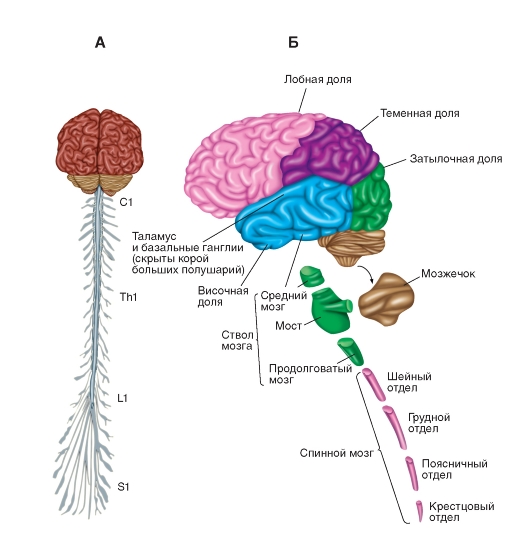
Small. Scheme of the central nervous system (central nervous system). dzherelo: Atlas of Physiology. Have two volumes. Volume 1: navch. posibnik / A.G. Kamkin, І. S. Kiselova - 2010 .-- 408 p. (Http: //vmede.org/sait/? Page = 7 & id = Fiziologiya_atlas_kamakin_2010 & menu = Fiz ...)
Video: Central nervous system
The nervous system in the functional and structural relationship lasts for peripheralі central the nervous system (CNS).
The central nervous system folds into headі dorsal brain.
The cerebrum is located in the middle of the cerebral cavity of the skull, and the spinal cord is in the spinal canal.
The peripheral part of the nervous system is composed of nerves, so that bundles of nerve fibers, which go beyond the boundaries of the brain and spinal cord and up to the other organs of the body. Before her, the same nerves are brought to the universities, for ganglia- the purchase of nerve cells in the position of the spinal cord and the brain.
The nervous system functions as a single unit.
Functions of the nervous system:
1) formulation of zbudzhennya;
2) transfer of zbudzhennya;
3) galmuvannya (reduction of damage, reduction of its intensity, reduction, reduction of expansion of growth);
4) integration (registration of new flows and changes of flows);
5) the removal of the development from the external and internal center of the body for the additional help of special nerve cells - receptors;
6) koduvannya, to reinterpret the chemical, physical teasing in the nerves of the impulse;
7) trophic, or lively, function - the establishment of biologically active speech (BAR).
neuron

the appointment of the witness
Neuron is the main structural and functional unit of the nervous system.
neuron
- the process of specializing the otrochtaya clitine, healthy sprymati, conducts and transmits the nerves for the processing of information in the nervous systems. © 2016 Sazonov V.F ..
Neuron - tse foldably vashtovanny itchy secreting highly differentiated nervova klitina with sprouts, Yaka spriymaє nerve zbudzhennya, reprobable yogo and transferring to our clients. Krym a wakeful injected neuron can press on its targets as well as modulating injections.
Robot galvanized synapse
Galmic synapse on its postsynaptic membrane MAC receptor to a galmic mediator - gamma-aminobutyric acid (GABA or GABA). At the end of the wakeful synapse in the halmic synapse, on the postsynaptic membranes, GABA displays the channel not for sodium, but for chlorine. They bring chlorine into the cell not a positive charge, but a negative one, to counteract the excitement, to neutralize the positive charge on sodium, to stimulate the cell.
Video:Robot of the GABA receptor and the halmic synapse
Otzhe, the destruction through the synapse is transmitted by a cheeky shlyah for the help of special cheruyuchy speeches,to be found in synaptic bulbs, roasted in presynaptic plaques... Zagalna is called tsikh rechovin - neurotransmition
, Tobto "Neurotransmitters". Їх subscribe tomediatori (mediators), which convey an awakening or a galmuvannya, i modulator I will change the position of the post-synaptic neuron, I will transfer it to the post-synaptic neuron.